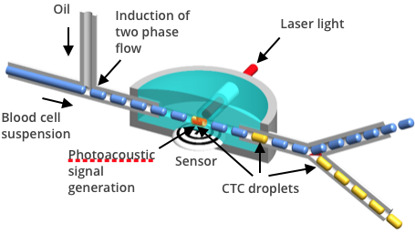
Photoacoustic detection of CTCs involves passing blood through a laser beam, which slightly heats cells, causing them to expand and produce a sound signal. Cancer cells are distinguished from normal cells by a unique sound signal they produce (melanoma), or by a sound signal they produce when tagged with a tumor directed monoclonal antibody linked to a sound producing particle, such as a light absorbing nanoparticle. The tumor cells remain viable through this process. The technology is applicable to detection of a broad range of cancers, including melanoma, breast, prostate, colon, lung, ovarian, colon, and other cancers.
Photoacoustic Detection and Capture of CTCs
Detection and Capture of Circulating Melanoma Cells (CTCs)
• Melanoma, a deadly skin cancer, is unique in that over 95% of melanomas are pigmented due to the presence of melanin.
• The presence of melanin in CTCs allows them to be detected by Acousys Biodevices’ photoacoustic method.
• A pulsed laser is flashed (in nanosecond bursts) at the blood buffy coat sample as it passes through a flow detection chamber.
• White blood cells do not generate a photoacoustic signal as they pass through the laser light, but naturally pigmented CTCs generate a robust acoustic wave.
• Once a CTC is detected by the production of a photoacoustic wave, it can be automatically separated from the blood sample using a microfluidic technique and captured for confirmation of identity and further analysis by various methods.
• The CTCS 3.0 can detect and capture a single CTC in a 10 mL blood sample.
• Typical processing time of the buffy coat from a 10 mL sample of blood requires ~16 minutes.
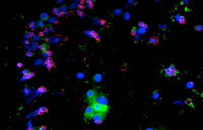
Fluorescently stained melanoma cells captured by photoacoustic method
Application to Other Types of Cancers
• Standardized tagging techniques enable the application of the photoacoustic detection process to other non-melanoma cancers using monoclonal antibodies.
• The monoclonal antibody tagging technique allows attachment of colored microspheres or nanoparticles to a non-pigmented cancer.
• Colored microspheres or metallic nanoparticles absorb laser light and subsequently generate high frequency ultrasonic waves, indicating the presence of CTCs.
• Photoacoustic detection can be used to detect CTCs of many other types of cancer including breast, prostate, lung, colon, ovarian, and other cancers.
• Captured CTCs can be immuno-fluorescently stained to verify their identity.
• The captured CTCs can be further studied by genetic and other methods.